Electron Microscopy
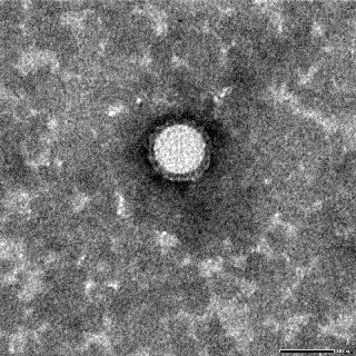
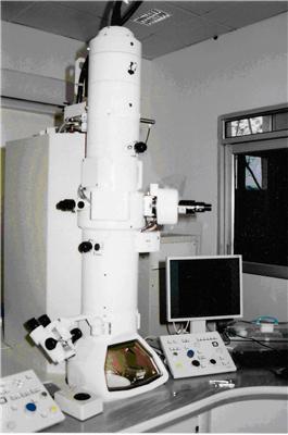
The possibility for high magnifications and greater resolutions has made the Transmission Electron Microscope (TEM) a valuable tool in both biological and materials research. A TEM facility costing about Rs. 1.80 crores is available in a dedicated building at NIHSAD campus. This was made possible through a special grant sanctioned by Hon’ble Director General ICAR, Dr. Mangla Rai during Xth plan after the first outbreak of bird flu (Avian influenza HPAI-H5N1) in 2006. The unit was inaugurated on 31.07.2009. EM unit of NIHSAD has a 120 kV class high-resolution transmission electron microscope (JEM-1400, Jeol, Japan). The unit also has an ultramicrotome (Ultracut-UCT, Leica) and a knife maker (EMKMR2, Leica) for ultra-thin sectioning of tissue samples. The services of the TEM unit add to the existing array of diagnostic facilities available at NIHSAD for quick confirmation of various exotic and emerging viral infections of animals. Apart from being useful in ultrastructural studies on viral infections, it can be of great help in identification and confirmation of viral etiology in case of unknown disease outbreaks.
Transmission Electron Microscope (TEM) Unit
Contact:
Dr. K. Rajukumar
Principal Scientist and In-charge Electron Microscopy, NIHSAD
email: k[dot]rajukumar(at)icar(dot)org(dot)inEqual Opportunity for Persons with Disabilities
In accordance with the provisions of the Right of Persons with Disabilities Act, 2016 and Rules, the institute strives to provide opportunities and facilities to persons with disabilities to participate, perform and excel in their work on an equal basis in everyday life.